Cortical-SSM: A Deep State-Space Model for EEG/ECoG Motor Imagery Decoding

Under Review

Currently, the paper is under review. We will set the links as soon as the paper is published.
At this moment, our code and additional report are provided as supplementary materials.

An overview of the MI classification task. The input consists of EEG/ECoG recorded while the subject imagines actions such as an elbow extension which should be predicted by the model.

Abstract

Classification of electroencephalogram (EEG) / electrocorticogram (ECoG) signals during motor-imagery (MI) exhibits significant application potential, including communication assistance and rehabilitation support for patients with motor impairments. These signals remain inherently susceptible to physiological artifacts (e.g., eye blinking, swallowing), posing persistent challenges.

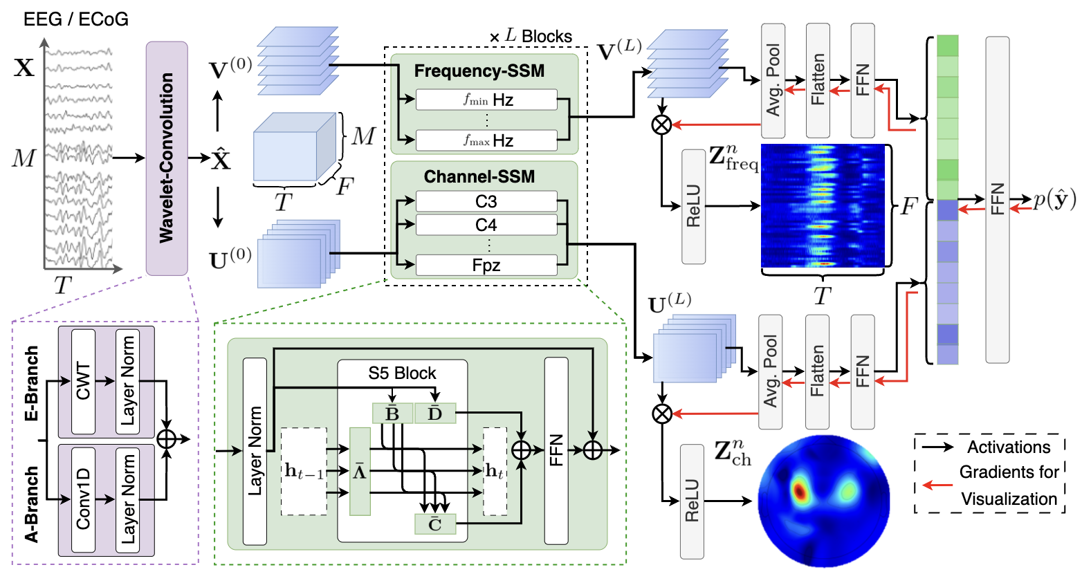
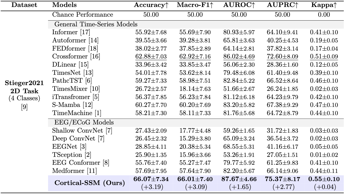
To overcome these limitations, we propose Cortical-SSM, a novel architecture that extends deep state-space models to capture integrated dependencies of EEG/ECoG signals across temporal, spatial, and frequency domains.

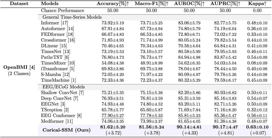
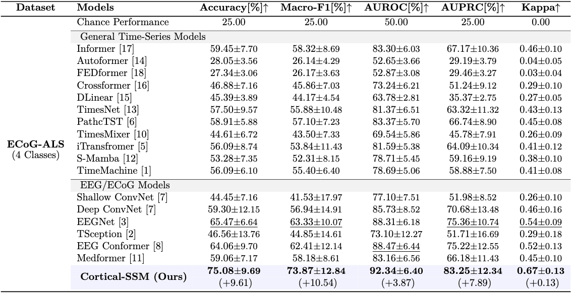
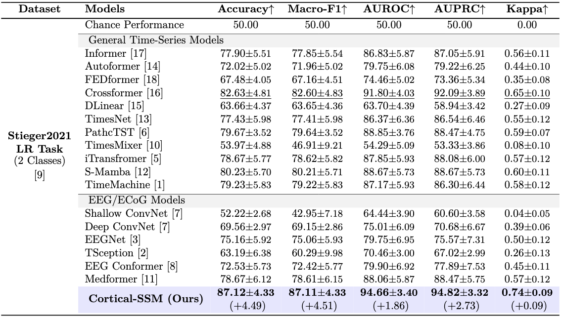
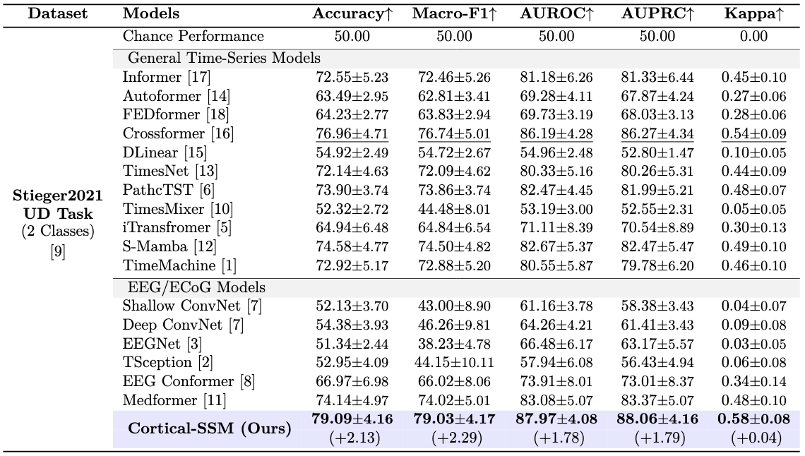
For evaluation, we conduct comprehensive evaluation across three benchmarks: 1) Two large-scale public MI EEG datasets containing >50 subjects, 2) A clinical MI ECoG dataset recorded from an amyotrophic lateral sclerosis patient. Across these three benchmarks, our method outperformed baseline methods on standard evaluation metrics. 
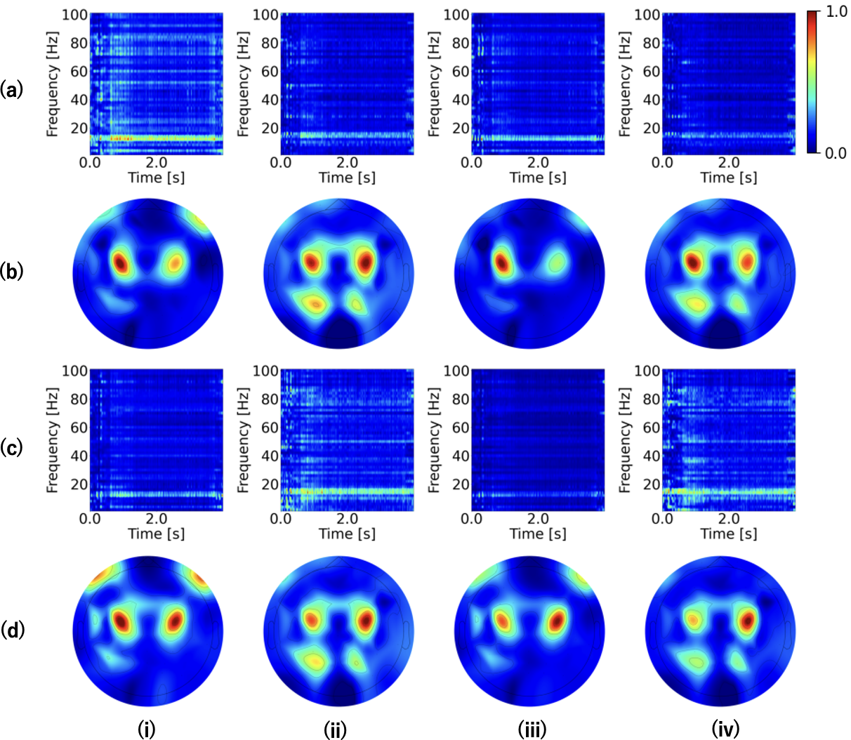
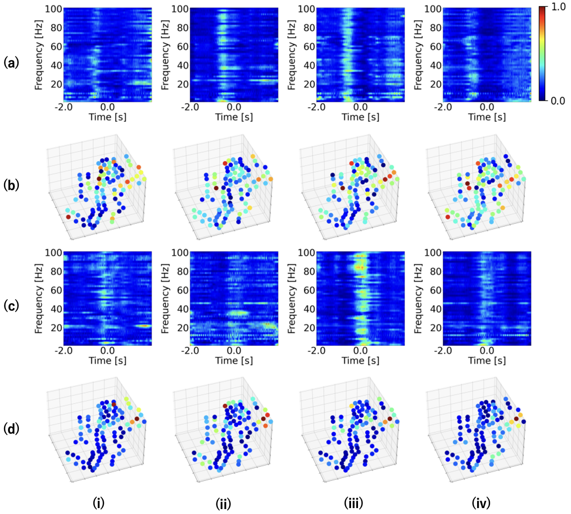
Furthermore, visual explanations derived from our model indicate that it effectively captures neurophysiologically significant regions in both EEG/ECoG signals.

Overview

  • 1. Wavelet-Convolution
    We introduce Wavelet-Convolution, which integrates deterministically obtained frequency components with adaptively derived frequency features. This integration enables the extraction of interpretable, non-black-box features while maintaining learnable representation.
  • 2. Frequency-SSM / Channel-SSM
    We extend Deep SSM by introducing Frequency-SSM, which captures spatio-temporal features for individual frequency components, and Channel-SSM, which captures temporal-frequency features for individual electrodes. This enables the modeling of integrated dependencies across temporal, spatial, and frequency domains.


.
Overview of the proposed Cortical-SSM.

Datasets

We evaluate our method on three datasets:

  • 1. OpenBMI (public EEG dataset)
    OpenBMI is a public dataset of EEG recorded from 54 healthy subjects during a MI task. The task involves two types of MI: right-hand grasping and left-hand grasping. Subjects engaged in 2 sessions, performing 400 MI trials per session, and a total of 21,600 samples were collected from 54 subjects.
  • 2. Stieger2021 (public EEG dataset)
    Stieger2021 is a public EEG dataset recorded from 62 healthy subjects, performing MI tasks involving cursor control. Three task types were used: LR (left vs. right-hand grasping), UD (bilateral grasping vs. rest), and 2D (combining LR and UD). Subjects completed 7–11 sessions, with 1,050–1,650 trials per task, totaling 269,099 samples. EEG signals were recorded at 1,000 Hz using 64 electrodes (10–10 system).
  • 3. ECoG-ALS (in-house ECoG dataset)
    ECoG-ALS is an electrocorticography (ECoG) dataset collected from a single patient with amyotrophic lateral sclerosis (ALS),who performed motor imagery (MI) tasks involving four movement types: elbow extension, elbow flexion, hand extension, and hand grasping. Each movement was repeated 40 times per session across eight sessions, resulting in a total of 1,280 samples. ECoG signals were recorded at a sampling rate of 1,000 Hz from 94 subdural electrodes placed near the left central sulcus.
.
Electrode distribution in ECoG-ALS. Electrodes 9-25 and 49-60, positioned within the precentral and postcentral gyrus, respectively, are located in the Hand Knob Area.

Results

Here, we show the results of the proposed method on OpenBMI and ECoG-ALS.

Interpretability

Sample-agnostic visual explanations of the proposed method on OpenBMI. Rows (a) and (c) display the temporal-frequency visual explanations for left and right hand grasp, respectively, while Rows (b) and (d) show the spatio-temporal visual explanations for left and right hand grasp, respectively. Columns (i)-(iv) correspond to the subjects: (i) 02, (ii) 11, (iii) 20, and (iv) 27.


Sample-agnostic visual explanations of the proposed method on ECoG-ALS. Rows (a) and (c) display the temporal-frequency visual explanations for Sessions 06 and 07, respectively, while Rows (b) and (d) show the spatio-temporal visual explanations for Sessions 06 and 07, respectively. Columns (i)–(iv) correspond to the output classes: (i) elbow extension, (ii) elbow flexion, (iii) hand extension, and (iv) hand grasp.


Quantitative Results


BibTeX


    To be appeared.